Summary
Modern linear ultrasound probes are built to produce very detailed images of structures close to the body surface. Because they use high-frequency sound waves, they are especially good at showing vessels, tendons, nerves, and other small or superficial structures with excellent clarity. In both human and veterinary medicine, linear probes are commonly used for vascular exams, musculoskeletal imaging, thyroid and breast scans, nerve blocks, superficial masses, and soft tissue imaging in small animals or horses.
Introduction
Ultrasound machines use different types of probes, also called transducers, depending on what part of the body needs to be scanned, and understanding what an ultrasound probe is helps explain why probe selection matters. Each probe shape and frequency range is designed for a particular job.
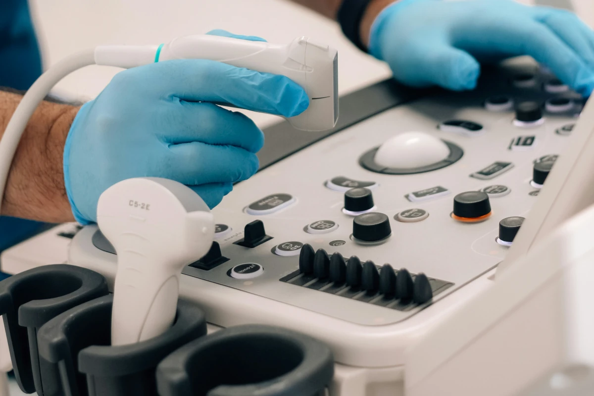
A linear probe has a flat face and a straight row of piezoelectric elements. That design creates parallel sound beams and produces a rectangular image with consistent detail across the scan. In simple terms, it is the probe you reach for when you need to see shallow structures clearly and in fine detail.
This article explains what a linear ultrasound probe is, where it is most useful, when another probe may be a better option, and how it is used in both human and veterinary imaging.
What Is a Linear Ultrasound Probe?
A linear probe is an ultrasound transducer with many small elements lined up in a straight row. Because the footprint is flat, the sound waves travel in parallel rather than spreading outward. That creates a rectangular image and helps maintain a more even field of view as depth increases.
Linear probes usually work at relatively high frequencies, often in the range of about 5 to 18 MHz. Higher frequency means better image detail, but it also means less depth penetration. So while linear probes are excellent for structures near the surface, they are not the best choice for deeper organs.
That is why they are commonly used for superficial anatomy such as blood vessels, tendons, nerves, skin, and small organs.
Main Uses of Linear Ultrasound Probes
Because linear probes give such clear detail near the surface, they are used in a wide range of exams.
They are especially common for:
Vascular imaging
Looking at arteries and veins, checking for clots, and guiding vascular access.
Musculoskeletal and soft tissue exams
Imaging tendons, ligaments, muscles, joints, and superficial masses.
Small parts imaging
Examining the thyroid, breast, testicles, lymph nodes, and other small superficial structures.
Needle guidance
Helping guide biopsies, injections, nerve blocks, fluid drainage, and similar procedures.
Veterinary soft tissue imaging
Scanning equine tendons and ligaments, small animal eyes, superficial masses, and certain abdominal structures.
Each of these uses comes down to the same strength: a linear probe gives high-resolution detail where depth is not the main challenge.
Vascular and Blood Vessel Imaging
One of the most common reasons to use a linear probe is vascular imaging.
Many arteries and veins sit fairly close to the skin, which makes them ideal targets for a high-frequency probe. The rectangular image helps the operator keep the vessel centered and follow it easily, while the strong near-field detail makes it easier to assess the vessel wall, surrounding tissue, and blood flow.
Linear probes are often used for:
Carotid and jugular imaging: Look for narrowing, plaque, or blockages in the neck vessels.
Peripheral vein assessment: Evaluate veins for thrombosis, mapping, or access planning.
Arterial exams: Assess diameter, wall changes, or superficial vascular disease.
Vascular access: Guide catheter placement in real time and confirm where the needle is going.
Because these probes also work well with Doppler, they are a strong choice when you need both anatomical detail and flow information.
Musculoskeletal and Soft Tissue Exams
Linear probes are also widely used for musculoskeletal imaging.
They are especially useful for tendons, ligaments, muscles, and superficial soft tissues because the high resolution helps show fine details such as fiber alignment, tears, swelling, fluid pockets, or changes in tissue structure.
In human medicine, they are commonly used for tendon injuries, ligament evaluation, joint-related soft tissue problems, and superficial masses. In veterinary medicine, they are especially important in equine tendon and ligament work.
Soft tissue imaging with a linear probe can also help identify things like abscesses, cysts, cellulitis, foreign bodies, or lipomas. Because the field of view stays fairly consistent, it is also useful when comparing the same area over time to monitor healing or treatment response.
Small Parts Imaging
Another major role for linear probes is small parts imaging.
These exams need very clear detail because the structures are small and often located close to the skin. Linear probes are a good fit for:
Thyroid ultrasound: Assess nodules, cysts, and gland structure.
Breast imaging: Evaluate superficial lesions and help characterize masses.
Testicular and scrotal exams: Assess torsion, tumors, varicoceles, or other abnormalities.
Lymph nodes and superficial glands: Check size, shape, and internal appearance.
Ophthalmic imaging: In some specialized settings, very high-frequency linear probes can be used for detailed imaging of the eye.
In all of these cases, the value of the linear probe comes from the same thing: high-resolution detail in superficial tissue.
Needle Guidance and Procedures
Linear probes are also extremely useful during ultrasound-guided procedures.
When placing a needle for a biopsy, fluid aspiration, injection, or nerve block, it helps to see both the needle and the target structure clearly. A linear probe makes that easier because its flat footprint and rectangular image are well suited to needle alignment, especially in superficial tissue.
These probes are commonly used for:
Nerve blocks: Guide anesthetic placement safely.
Biopsies: Target tissue more accurately.
Fluid drainage: Guide needle placement into a fluid pocket.
Vascular access: Improve success and reduce complications.
For many of these procedures, the ability to visualize the needle in real time is one of the main reasons a linear probe is chosen.
Veterinary Uses of Linear Probes
Linear probes are especially important in veterinary ultrasound because many common exams involve superficial soft tissues.
Equine applications
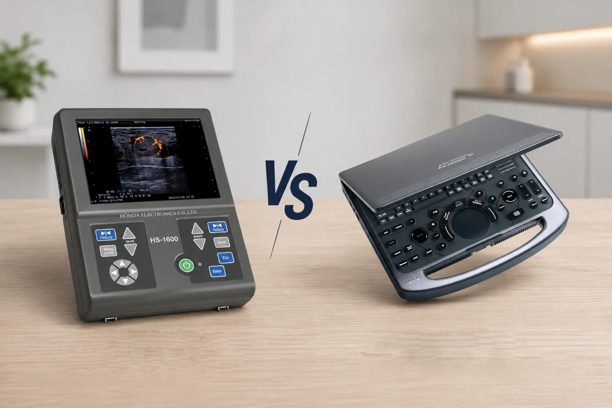
In horses, linear probes are widely used for tendon and ligament imaging. They help evaluate fiber pattern, lesion size, swelling, and healing progress, which makes them a key tool in sports medicine and rehabilitation, particularly when used with portable field systems such as Honda ultrasound systems in equine practice.
They are also useful for soft tissue swelling around joints, bursae, and ligaments. Because many equine scans happen in the field, portable ultrasound machines paired with linear probes are especially common.
Small animal applications
In dogs and cats, linear probes are often used for superficial abdominal structures, small organs, lymph nodes, eyes, and superficial masses. In cats in particular, the smaller body size can make a linear probe useful for structures that might sit deeper in larger animals.
They are also commonly used in ocular imaging and for soft tissue procedures.
Other veterinary uses
Veterinarians may also use linear probes for vascular access, skin masses, wound monitoring, biopsy guidance, and scans in exotic or very small patients. In those cases, smaller versions such as hockey-stick probes can be especially helpful.
Linear Probe vs Other Ultrasound Probes
A linear probe is very useful, but it is not the right tool for every job, which is why understanding linear vs convex ultrasound probes can help clinicians choose the best transducer for each exam.
Linear probe
A linear probe has a flat footprint, high frequency, and a rectangular field of view. It is best for superficial structures where image detail matters most.
Convex or curvilinear probe
A convex probe has a curved footprint and lower frequency. It produces a wider field of view and reaches deeper, which makes it better for abdominal imaging, obstetrics, and many general scans.
Phased-array probe
A phased-array probe has a small footprint and creates a sector-shaped image. It is especially useful for cardiac imaging because it can fit between the ribs and still reach deeper structures.
So while the linear probe is excellent for surface detail, other probes are better when the anatomy is deeper or harder to access.
When a Linear Probe Is Not the Best Choice
Despite its strengths, a linear probe is not always the right option.
Because it uses high-frequency sound, its depth penetration is limited. That makes it less useful for:
- Deep abdominal imaging
- Pelvic structures in larger patients
- Many cardiac exams
- Situations where bone or gas blocks the beam
If the target is too deep, or if the scan needs to go through the ribs or around a lot of bowel gas, a lower-frequency convex or phased-array probe is usually a better choice.
In general, the linear probe works best when the structure you care about is close enough to the surface for the high-resolution benefit to matter.
Conclusion
Linear ultrasound probes are one of the most important tools in diagnostic imaging when superficial detail is the priority. Their flat footprint and high-frequency design make them especially useful for blood vessels, tendons, ligaments, nerves, thyroid tissue, breast imaging, small superficial organs, and ultrasound-guided procedures.
In veterinary medicine, they are particularly valuable for equine tendon work, small animal soft tissue imaging, ocular scans, and superficial masses. In human medicine, they are a standard choice for vascular, musculoskeletal, and small-parts imaging.
They are not the best fit for every exam, especially when greater depth is needed, but for surface structures and fine detail, a linear probe is often the best tool available.