Summary
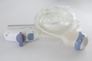
An ultrasound probe, often called a transducer, is the handheld device that sends and receives high-frequency sound waves during an ultrasound exam. It’s the part of the system that makes real-time imaging possible. In this guide, you’ll learn what a probe is, how it works, the main probe types, and how to choose and care for one.
Introduction
Ultrasound is a safe, non-invasive imaging method that uses sound waves, not radiation, to create real-time views of internal structures. It’s used every day in obstetrics, cardiology, emergency care, musculoskeletal imaging, and veterinary medicine.
At the heart of every scan is the probe (transducer). It delivers sound into the body and “listens” for echoes coming back. No probe, no image.
Knowing how probes differ is more than technical trivia. The right probe can sharpen image quality, improve diagnostic confidence, and speed up workflow. The wrong one can limit depth, reduce detail, or make scanning harder than it needs to be.
What Is an Ultrasound Probe?
An ultrasound probe is a medical device that produces sound waves above human hearing, typically in the megahertz (MHz) range, and detects the echoes that return from inside the body.
Most probes are used externally on the skin with a water-based gel. The gel removes the air gap between probe and skin so sound can travel efficiently. Some probes are designed for internal imaging, allowing the transducer to sit closer to the target organ for clearer detail.
Inside the probe are piezoelectric crystals. These crystals convert electrical energy into mechanical vibrations (sound waves), then convert returning vibrations back into electrical signals. That back-and-forth conversion is what makes ultrasound imaging work.
How Does an Ultrasound Probe Work?
Here’s the basic chain of events:
- The ultrasound system sends electrical pulses to the probe.
- Piezoelectric elements vibrate in response.
- Those vibrations create high-frequency sound waves.
- Sound waves travel through tissue and reflect off structures differently.
- Returning echoes make the crystals vibrate again.
- The probe converts those vibrations into electrical signals.
- The system measures echo timing and strength to build a real-time image.
The key factor is time. By measuring how long echoes take to return and how strong they are, the system estimates depth and helps distinguish tissue characteristics.
Types of Ultrasound Probes
Different exams require different probe designs. What matters most is frequency, footprint shape, and array configuration.
Linear Probes
Linear probes have a flat surface and usually run at higher frequencies (often 7 to 15 MHz). They’re built for crisp detail in superficial anatomy.
Common uses include:
- Vascular imaging
- Thyroid exams
- Musculoskeletal imaging
- Nerve blocks
They generate a rectangular image and are ideal when you need high-resolution views close to the skin.
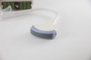
Curvilinear (Convex) Probes
Convex probes have a curved footprint that widens the field of view. They typically operate at lower frequencies (often 2 to 5 MHz), which improves penetration for deeper structures.
Common uses include:
- Abdominal imaging
- Obstetric exams
- Pelvic studies
Their curved design creates a broad sector-shaped image that’s well suited for larger internal organs.
Phased Array Probes
Phased array probes have a small footprint and use electronically timed activation to steer the ultrasound beam.
They’re ideal for:
- Cardiac imaging
- Intercostal scanning
- Tight access areas (between ribs)
Because the beam can be steered electronically, they’re especially helpful for imaging the heart through limited rib spaces.
Endocavitary Probes
Endocavitary probes are designed for internal exams, such as:
- Transvaginal imaging
- Transrectal imaging
By placing the probe closer to the anatomy of interest, they can provide higher detail and better resolution than external scanning in many cases. These probes also require strict cleaning and high-level disinfection between uses.
Specialty Probes (3D/4D, TEE, Volume)
Some clinical needs require advanced probe designs:
- 3D/4D probes: volumetric imaging, commonly in obstetrics
- TEE probes: inserted into the esophagus for detailed cardiac views
- Volumetric probes: capture large data sets for reconstruction and advanced analysis
These probes may include extra sensors or mechanical components to support specialized imaging modes.
Choosing the Right Probe
A good probe choice usually comes down to three things:
1) Frequency
- Higher frequency: sharper detail, less penetration
- Lower frequency: deeper penetration, less fine detail
If the target is superficial, go higher. If you need depth (abdomen, deep pelvis), go lower.
2) Depth
You can adjust depth on the ultrasound machine, but physics still applies: probe frequency influences how far sound can travel and return usable echoes. Deep targets generally require lower-frequency probes.
3) Array Shape and Footprint
- Linear: rectangular image, excellent near-field detail
- Convex: wider sector view for larger/deeper anatomy
- Phased: wedge-shaped view, small footprint for cardiac and rib spaces
Quick matching guide:
- Linear: vascular, MSK, thyroid, procedures
- Convex: abdominal, obstetric, general deep scanning
- Phased: cardiac, intercostal windows
Also confirm probe compatibility with your ultrasound system, and consider ergonomics, cable durability, and handling comfort, especially in busy clinical environments. For clinics relying on mobile imaging, access to reliable portable ultrasound depot repair can minimize downtime and keep workflow disruptions to a minimum.
Care and Maintenance
Good probe care protects patients and extends equipment life.
Start with pre-cleaning immediately after use to remove gel and organic material. Even if you used a probe cover, cleaning is still required.
Best practice is to follow your facility protocol, manufacturer instructions, and professional guidelines. In general:
- Clean after every exam using approved solutions or wipes
- Use high-level disinfection for internal probes
- Store probes to prevent cable strain and impact damage
Poor cleaning shortens probe lifespan and increases infection-control risk. When damage does occur, professional ultrasound probe repair services can often restore functionality and extend the life of the transducer, helping facilities avoid premature replacement costs. Proper storage matters too, especially for cables and connectors.
Conclusion
An ultrasound probe (transducer) is the essential component that sends and receives ultrasound waves to create real-time images. Each probe type is designed for specific clinical tasks, and selecting the right one means balancing frequency, depth, and footprint.
When clinicians understand how probes work, how they differ, and how to care for them correctly, imaging quality improves and equipment lasts longer, which supports better diagnostic outcomes.
Frequently Asked Questions (FAQ)
1. What is the most important part of an ultrasound probe?
The piezoelectric crystals. They convert electrical energy into sound waves and convert returning echoes back into electrical signals.
2. Why are there different types of probes?
Different anatomy requires different frequencies, footprints, and fields of view to balance resolution and penetration.
3. Can probes be used internally?
Yes. Endocavitary probes (transvaginal, transrectal) are designed for internal imaging to improve proximity and detail.
4. How should I clean an ultrasound probe?
Remove gel and organic material first using approved cleaning methods, then disinfect appropriately. Internal probes require high-level disinfection.
5. How do I choose the right probe for my exam?
Match the probe to the target depth and required detail. Higher frequency gives sharper resolution at shallow depths; lower frequency penetrates deeper with less detail.