
Summary
Linear and convex (curvilinear) ultrasound probes mainly differ in frequency, imaging depth, and field of view. Linear probes run at higher frequencies, giving sharp detail for superficial structures. Convex probes use lower frequencies, letting you see deeper anatomy with a wider view. The “best” probe depends on what you’re scanning and what you need to answer clinically.
Introduction
Ultrasound probes (also called transducers) are the handheld part of the system that sends and receives sound waves to create live `images. The machine does the heavy processing, but the probe choice affects what kind of image you can get in the first place.
Because different probes are built for different jobs, knowing the difference between linear and convex probes helps you pick the right one, dial in image quality faster, and improve diagnostic confidence. If your current probe is showing signal dropouts, cable wear, or element failure, professional ultrasound probe repair services can often restore performance without the cost of full replacement.
In this guide, we’ll break down what each probe is, how they compare, where each one shines, and how to choose quickly in real-world scanning.
What Is a Linear Probe?
A linear ultrasound probe has a flat, rectangular face with a straight row (or rows) of piezoelectric elements. It creates a rectangular image with scan lines that stay parallel.
Linear probes run at high frequencies, often above 12 MHz. Higher frequency means shorter wavelengths, which gives excellent resolution, but it also means the sound waves don’t travel as far. So you get great detail, just not at deep depths.
Common uses for linear probes include:
- Vascular access (IVs, central lines)
- Musculoskeletal imaging (tendons, ligaments, joints)
- Soft tissue checks (lumps, abscess vs cellulitis)
- Thyroid exams
- Superficial nerves and nerve blocks
- Breast imaging
In short: if the target is close to the skin and you need fine detail, linear is usually your go-to. For deep abdomen or pelvis, it’s generally not the best match.
What Is a Convex (Curvilinear) Probe?
A convex probe (often called curvilinear) has a curved footprint. That curved shape produces a fan-shaped or trapezoidal image that widens as depth increases, which is perfect when you want to see a broader area inside the body.
Convex probes run at low to mid frequencies, commonly around 2–5 MHz. Lower frequency penetrates deeper, but the tradeoff is less crisp detail compared to a high-frequency linear probe.
Common uses for convex probes include:
- Abdominal imaging (liver, gallbladder, spleen, kidneys)
- Obstetric and gynecologic scans
- Trauma exams (FAST)
- Aorta and renal assessments
- Deep organ evaluation in larger patients
In short: if you need depth and a wide view of large anatomy, the convex probe is the workhorse.
Key Differences Between Linear and Convex Probes
1) Frequency and Image Quality
Linear probes:
- High frequency (often >12 MHz)
- Excellent resolution
- Limited depth
Convex probes:
- Low to mid frequency (often 2–5 MHz)
- Deeper penetration
- Moderate resolution
A helpful way to remember it: higher frequency = sharper image, shallower reach.
2) Field of View and Footprint
Linear probes:
- Flat footprint
- Rectangular field of view
- Great for lining up with vessels, tendons, and nerves
Convex probes:
- Curved footprint
- Wide, expanding field of view with depth
- Great for surveying large areas (abdomen, pelvis, pregnancy)
3) Typical Applications
Linear probes excel at:
- Vascular access
- MSK and tendon imaging
- Thyroid and breast imaging
- Nerve blocks
Convex probes excel at:
- Abdominal organ imaging
- OB/GYN scans
- FAST exams
- Deep structure assessment
Advantages and Limitations
Linear Probe
Advantages:
- Very high resolution
- Precise imaging for superficial anatomy
- Excellent for needle guidance and procedures
Limitations:
- Doesn’t penetrate well at depth
- Smaller overall field of view
- Image quality is sensitive to alignment and probe angle
Convex Probe
Advantages:
- Strong depth penetration
- Wide field of view for big anatomy
- Ideal for abdominal and OB scanning
Limitations:
- Lower resolution for superficial detail
- Not the best choice for fine structures near the skin
- Can show more distortion at very shallow depths
How to Choose Between Linear and Convex Probes
A quick decision comes down to three questions:
- How deep is the target?
- How much detail do I need?
- What’s the clinical task (survey vs precision vs procedure)?
Choose a linear probe when:
- You’re scanning superficial structures
- You need crisp detail
- You’re guiding a needle (lines, blocks, drainage)
Choose a convex probe when:
- You’re scanning deep organs
- You need a wide view
- You’re doing abdomen, OB/GYN, FAST, or aorta checks
In practice, lots of clinicians use both in one exam: convex to survey deep anatomy, then linear to zoom in on superficial findings or guide a procedure.
Also consider: patient body habitus, clinical setting (ER vs clinic), and probe compatibility with your ultrasound system. For practices relying heavily on portable units, scheduled evaluations through a structured portable ultrasound depot repair program can help ensure probes, boards, and imaging components stay within manufacturer specifications.
Popular Brands and Where to Buy
Most major ultrasound manufacturers offer both linear and convex probes across portable and cart-based systems.
For rugged, field-ready veterinary imaging, explore Dramiński portable ultrasound systems designed for demanding environments and interchangeable probe configurations.
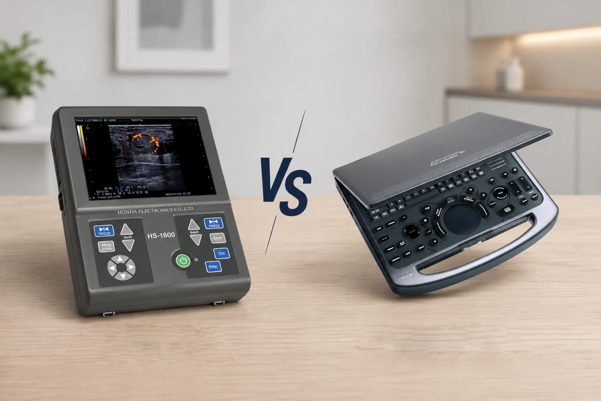
For dependable portable imaging in medical and veterinary settings, see Honda ultrasound systems for sale, known for consistent image quality and durable hardware.
When choosing a probe or system, prioritize compatibility, support, and long-term serviceability.
Conclusion
Linear and convex ultrasound probes are built for different jobs. Linear probes give high-resolution detail for superficial structures, while convex probes provide better depth and a wider view for abdominal and obstetric scanning.
Neither is “better” overall. The best probe is the one that matches the patient, anatomy, and clinical question. Many practices get the most flexibility by having both available.
Frequently Asked Questions (FAQ)
1. What frequency ranges do linear and convex probes use?
Linear probes usually operate above 12 MHz for high-detail superficial imaging. Convex probes commonly use 2–5 MHz for deeper penetration.
2. Which probe is better for vascular access?
Typically linear, because it shows superficial vessels more clearly and supports precise needle guidance.
3. Can I use a convex probe for musculoskeletal imaging?
You can, but it’s usually not ideal. MSK structures are often superficial, and linear provides the detail you want.
4. Why is resolution different between linear and convex probes?
Resolution improves as wavelength gets shorter. High frequency (linear) means shorter wavelength and better detail. Low frequency (convex) travels deeper but sacrifices sharpness.
5. Do I need both probes?
Many clinics do. Linear covers superficial detail and procedures, while convex handles deep organs and broad surveys. Your scope and patient population determine what’s essential.