Summary
Veterinary ultrasound courses usually combine lecture-based learning with plenty of hands-on scanning. Some are short, single-day sessions held in a clinic, while others are multi-day workshops covering everything from basic abdominal imaging to advanced echocardiography. The best course for you will depend on your current experience and what you want to use ultrasound for in practice.
Introduction
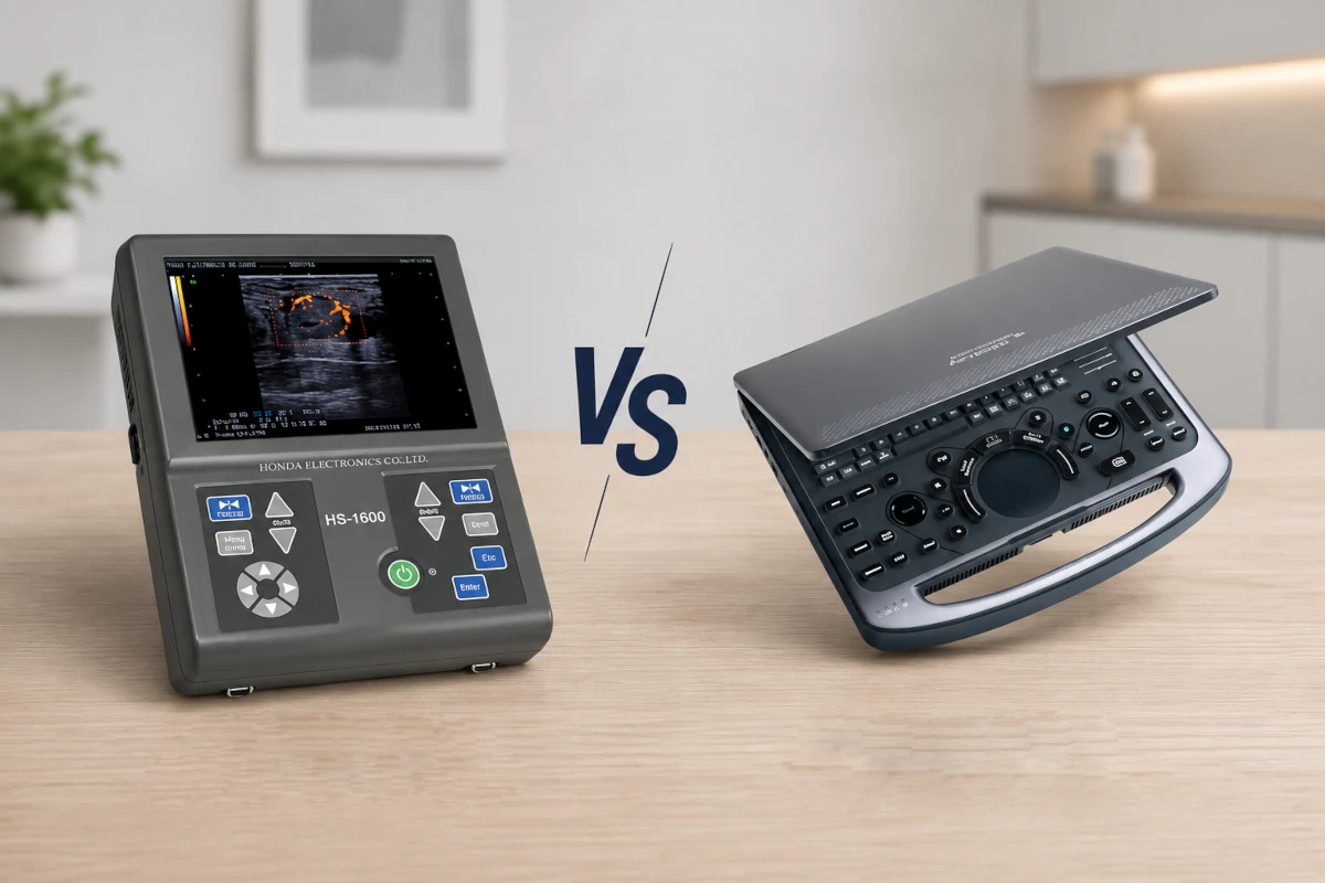
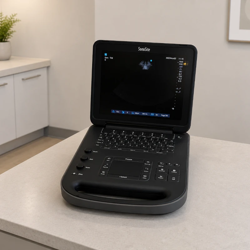
Point-of-care ultrasound, or POCUS, has become an essential part of modern veterinary medicine, supported by advances in portable ultrasound machines types that make bedside and in-clinic training more accessible. It helps clinicians quickly evaluate organs, detect free fluid or air, and guide procedures with greater accuracy. Formal training can improve diagnostic confidence, reduce the need for referrals, and ultimately support better patient care.
As ultrasound becomes more common in everyday practice, more veterinarians and nurses are looking for courses that offer both solid theory and meaningful scanning time. This guide explains how training programs are typically structured, from the basics through advanced abdominal and cardiac techniques, so you can choose the right option for your team, especially when considering structured options like veterinary ultrasound training designed for different experience levels.
Course Structure and Levels
Most training providers organise their courses in stages, starting with the basics and building toward more advanced abdominal work and specialised cardiac imaging.
Level I: Ultrasound Fundamentals
Introductory courses are designed for people with little or no prior ultrasound experience. They usually cover basic terminology, patient preparation, and core scanning principles.
Common topics include machine controls, image display modes, recognising artefacts, and understanding image orientation, along with practical concepts such as linear vs convex ultrasound probes and when to use each in clinical scanning. These courses often also introduce Doppler modes, ultrasound-guided fine-needle aspirates and biopsies, and a general survey of the abdominal organs, along with basic thoracic and cardiac views.
Level I is a good fit for veterinarians or technicians who are new to ultrasound.
Level II: Advanced Abdominal Ultrasound
Once you are comfortable with the basics, Level II courses move into a more structured abdominal protocol. Participants learn how to carry out rapid abdominal surveys as well as more detailed assessments of the liver, urinary bladder, kidneys, adrenal glands, pancreas, gastrointestinal tract, and reproductive organs.
These courses often recommend setting aside 6 to 8 hours each month for practice. In most cases, attendees are expected to have completed a basic course or be able to show equivalent experience.
Advanced Echocardiography
Cardiac ultrasound courses usually build on prior abdominal ultrasound experience. Participants learn proper patient positioning using right and left parasternal windows, how to obtain long-axis and short-axis views, perform M-mode measurements, and evaluate cardiac function.
The curriculum typically includes pulse wave, continuous wave, and colour flow Doppler. As with advanced abdominal courses, providers often recommend 6 to 8 hours of practice each month and expect attendees to have completed basic ultrasound training first.
Hands-On Learning and Course Logistics
Good ultrasound training balances lectures with substantial lab time.
For example, the Viticus Group’s two-day introductory abdominal course in Las Vegas combines detailed teaching on machine operation and image optimisation with hands-on scanning of dogs and cats. The course keeps the student-to-instructor ratio low and includes about 8 hours of lab work so participants can build confidence in obtaining diagnostic images and recognising normal anatomy. The program offers 16 CE hours, costs US $2,850, and is capped at 45 participants.
Other providers offer shorter, more focused formats. FASTVet’s Global FAST® program combines lectures and hands-on scanning into a single day, providing 9 hours of RACE-approved CE. These courses usually run from 8:00 AM to 6:00 PM and follow case-based protocols such as AFAST, TFAST, and Vet BLUE. Each scanning station typically includes three to four attendees and one trained instructor, allowing for direct feedback throughout the day. Participants also receive course manuals and a certificate of completion.
For those wanting a more intensive experience, UniVets Global’s Ultrasound Academy offers five days of fully hands-on training with a dedicated tutor. Students work with different machines to become familiar with a range of systems and can earn VetCEE credits. The focus is on continuous scanning, individual feedback, and a supportive setting where attendees can concentrate on improving their skills.
Custom and In-Clinic Training Options
Some providers offer on-site training tailored to your clinic. These full-day sessions are usually limited to one to three staff members and focus on machine optimisation, image acquisition, and real-time feedback while scanning live patients.
These sessions typically last around 8 hours, are often scheduled on weekends, and may count toward continuing education credits. Requirements usually include an exam room, a working ultrasound machine, and access to five healthy animals that are fasted and shaved.
Sedation may be needed for anxious or fractious patients, especially if ultrasound-guided biopsies are part of the session.
Preparing for Training
Pre-course Knowledge
A bit of preparation beforehand can make a big difference, especially for Level II or advanced courses. Reviewing ultrasound physics and abdominal anatomy is often helpful, and many providers require participants to complete a basic course before moving on to advanced material.
It is also worth reading the course agenda ahead of time and refreshing your knowledge of the organs you will be scanning.
Physical Preparation
Wear comfortable scrubs and closed-toe shoes, since hands-on labs usually involve standing at a scanning table for long periods. If you are travelling to a course, it can help to bring a sweater for cold lecture rooms and sunscreen for outdoor breaks.
If you are bringing your own machine, make sure it has been serviced and that you have the appropriate probes.
Animal Preparation
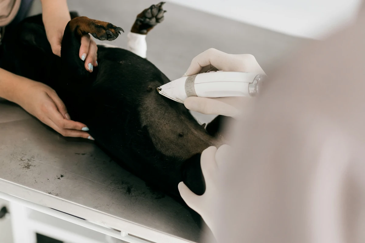
When live animals are used, they are usually fasted overnight and shaved over the abdomen to improve probe contact and image quality. Sedation is not commonly needed for routine abdominal scans, but it may be used for anxious or aggressive animals and is often necessary for ultrasound-guided biopsies.
Animals should be stable before sedation, and drug choices should be adjusted based on age, temperament, and medical history.
Equipment
If you need to bring your own ultrasound machine, check that both the unit and probes are working properly, and reviewing what an ultrasound probe is can help reinforce how probe condition affects image quality during training. Instructors often help participants optimise their own equipment during the course, so it is also a good idea to bring any accessories you regularly use, such as needle guides or transducer covers.
Choosing the Right Training Program
The right course depends on your experience level and your clinical goals. Beginners are usually best served by Level I or basic abdominal training, while those with more experience may be ready for Level II abdominal courses or specialised echocardiography training.
When comparing programs, consider the following:
Lecture versus hands-on time
Look for courses that offer substantial lab time, ideally 8 to 16 hours, along with a low student-to-instructor ratio.
Instructor qualifications
Programs taught by board-certified radiologists or experienced sonographers can offer stronger clinical guidance. FASTVet’s courses, for example, are led by Dr. Gregory Lisciandro, a widely recognised author and educator.
Class size and feedback
Smaller groups usually allow for more personalised instruction, which can make a big difference when developing scanning skills.
Continuing education and cost
Check both CE hours and tuition. The Viticus Group’s basic course offers 16 hours for $2,850, but fees and course length vary across providers.
Location and convenience
Choose a format that works for your practice. In-clinic training can be especially useful for teams that want focused instruction without the need to travel.
Conclusion
Veterinary ultrasound training usually starts with the fundamentals of physics, machine controls, and image orientation before moving into more advanced abdominal protocols and echocardiography. Whether the course lasts one day or several, the most effective programs combine teaching with plenty of hands-on scanning.
Preparing in advance by reviewing the basics, dressing appropriately, and making sure your equipment and animals are ready can help you get more out of the experience. By choosing a course that matches your current skill level and clinical goals, and by committing to regular practice, you can improve diagnostic confidence, support better patient care, and get more value from your ultrasound equipment.
Frequently Asked Questions (FAQ)
How long do veterinary ultrasound training courses last?
Course length varies. Basic abdominal courses often run for two days, offering 16 CE hours and around 8 hours of lab time. FASTVet’s Global FAST program fits lecture and scanning into a single 9 to 11 hour day. Some academies also offer five-day intensive programs with continuous practice.
Do I need prior ultrasound experience?
Not always. Introductory courses are designed for participants with little or no previous experience. Advanced abdominal and echocardiography courses usually require completion of a basic course or similar prior knowledge.
What will I learn during training?
You can expect to learn how to use machine controls, improve image quality, identify normal anatomy such as the liver, spleen, kidneys, adrenal glands, pancreas, bladder, and gastrointestinal tract, and perform systematic scanning routines. Advanced courses focus on organ-specific protocols and echocardiographic techniques.
Are animals used during hands-on sessions?
Yes. Most courses include scanning live dogs and cats. In-clinic sessions may require five healthy, fasted, shaved animals, and sedation may be used for anxious pets or invasive biopsy procedures.
Is sedation always required?
No. Sedation is not usually needed for routine abdominal ultrasound. However, it may be used for animals that are difficult to handle and is often required for ultrasound-guided biopsies to improve safety and image quality.
What equipment do I need to bring?
Most participants use the machines provided by the course. For in-clinic training, however, you will usually need to provide a working ultrasound system and the appropriate probes. Comfortable clothing, closed-toe shoes, and any accessories relevant to your scanning protocols are also recommended.