Summary
Veterinary ultrasound is no longer limited to referral centers. In many general practices, it is now part of everyday clinical decision-making because it offers real-time imaging, can be repeated easily, and supports rapid bedside assessment in both routine and emergency care. POCUS protocols are designed to answer focused clinical questions and can often be completed in under 10 minutes, which makes them especially useful for triage and ongoing monitoring.
This guide explains how to use ultrasound in a practical, patient-friendly way without changing the core principles of good medicine. It covers equipment, training, patient preparation, scanning technique, sedation, image interpretation, ultrasound-guided sampling, and the small details that make the experience easier for both patients and owners.
Introduction
For modern veterinary teams, ultrasound has become a practical extension of the physical exam. Portable units now make it possible to perform point-of-care ultrasound in the consult room, treatment area, or field, giving clinicians a fast way to look for free fluid, assess organs, and support immediate treatment decisions. These focused exams are not a substitute for a full specialist study, but they are valuable because they are repeatable, objective, and designed to answer clear clinical questions in real time.
Compared with radiography, ultrasound uses sound waves rather than ionizing radiation, making it a safe option for repeated imaging when clinical monitoring is needed. As more practitioners adopt POCUS, training remains the difference between simply owning a scanner and using it well. A 2024 study of 113 veterinary participants found that even a short, structured two-day course significantly improved confidence across abdominal, pleural-lung, cardiac, and ultrasound-guided vascular access applications.
Equipment and Training
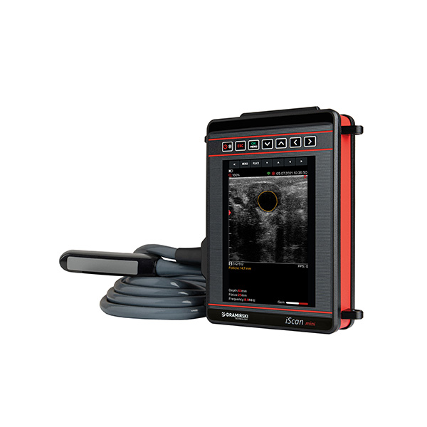
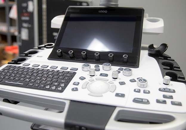
Veterinary practices now have access to a wide range of ultrasound systems, from full cart-based machines to handheld devices, and clinicians comparing options may benefit from this veterinarian’s guide to choosing the right ultrasound machine when evaluating systems for their practice. Handheld platforms have widened access to ultrasound in general practice and emergency settings, while larger machines still offer stronger image optimization, Doppler options, and workflow flexibility for more advanced studies.
Probe choice matters, and understanding the differences between transducer designs—such as those explained in linear vs convex ultrasound probes, can help clinicians select the most appropriate probe for each exam. Linear probes provide high-frequency detail for superficial structures and musculoskeletal work. Curvilinear or convex probes are often better for abdominal surveys because they balance depth and field of view. Phased-array probes are useful for cardiac imaging because they fit narrow acoustic windows. BMUS guidance recommends using the highest frequency that still provides enough depth, since this improves resolution without sacrificing penetration.
Training matters just as much as equipment, which is why many clinicians pursue structured programs such as veterinary ultrasound training to develop consistent scanning technique and interpretation skills. Good ultrasound takes more than knowing the controls. Clinicians need a systematic method, an understanding of normal anatomy and artifacts, and the judgment to know when a focused scan is enough and when referral is the better choice. Hands-on teaching and repetition make a real difference, and current evidence supports structured training as a practical way to increase confidence and consistency.
Patient Preparation
Fasting and bladder preparation
Preparation improves image quality, but it should also stay practical. For scheduled abdominal ultrasound, BMUS recommends fasting for ideally a minimum of 8 hours, with water allowed, because this can improve visualization of the gastrointestinal tract and reduce interference from luminal contents.
That said, fasting is helpful rather than absolute in every case. A prospective study of 150 dogs found that routine fasting was not essential for abdominal ultrasonography, which is a useful nuance when urgent or same-day imaging is needed. In practice, fasting still makes sense for many routine abdominal exams, but clinicians should balance ideal preparation with the patient’s condition and the urgency of the scan.
For urinary and reproductive studies, a moderately filled bladder is ideal. BMUS notes that bladder filling affects image quality and that the bladder is best imaged when moderately full. For owners, that usually means avoiding a final toilet break right before arrival when a bladder study is planned.
Clipping and skin contact
Ultrasound depends on good contact. Air blocks the beam, so clipping is still the best option for most diagnostic scans. In abdominal studies, this usually means clipping a broad enough window to avoid constantly chasing anatomy at the edge of the prep area. Generous gel should then be applied to remove trapped air and improve contact.
In some short-haired, unstable, or stress-sensitive patients, wetting the coat heavily with gel may be an acceptable compromise, but image quality may be lower. Endocavitary work is different and requires probe covers, lubricant, and proper disinfection protocols between patients.
Environment and comfort
A calmer patient usually means a better, safer scan. A quiet room, gentle handling, non-slip bedding, and warm gel all help reduce tension and make the process feel less clinical and more manageable for the animal. Fear Free guidance emphasizes that ultrasound can be integrated in a way that is more comfortable for both patients and owners, especially when expectations and handling are thoughtful from the start.
Owners can help too. Pets that are conditioned to gentle restraint, side-lying, standing still, or tolerating touch to the abdomen often scan more smoothly in clinic. That kind of cooperative care does not replace restraint when needed, but it can reduce the amount of restraint required and improve the overall experience. This is particularly valuable for repeat imaging in chronic disease or follow-up visits.
Sedation and Fear Reduction
Sedation is not automatically required for ultrasound. Many abdominal and cardiac scans can be completed on conscious patients with gentle handling and good positioning. In fact, one of the advantages of ultrasound-guided fine-needle aspiration is that it usually requires very little to no sedation.
Still, sedation can make the difference between a rushed, frustrating scan and a complete diagnostic exam. Anxious, painful, boisterous, or reactive patients often benefit from light sedation or anxiolysis, especially when clinicians need detailed images or when emergency POCUS is being used to answer time-sensitive questions. Fear Free guidance also supports explaining the process, expected findings, and likely next steps to owners beforehand, since better owner understanding often improves compliance and lowers stress around the procedure.
Drug choice should always be tailored to the patient’s age, temperament, clinical status, and goals for the scan. There is no single protocol that fits every animal. For tissue sampling, the level of sedation depends on invasiveness: FNA often needs little or no sedation, while core biopsy is more invasive and is usually performed under heavy sedation or anesthesia because the complication risk is higher.
Positioning and Restraint
Positioning should support both image quality and patient safety. Small animal abdominal scans are commonly performed in lateral recumbency, but ventrodorsal positioning can improve access to some regions when the patient tolerates it. BMUS notes that there is no single right or wrong position, and that scan position should be guided by both clinical presentation and the operator’s ability to examine the abdomen thoroughly.
Patients with respiratory compromise, pleural effusion, or severe distress should never be forced into a position that worsens breathing. Some studies can be done standing, and in large dogs that may be the least stressful and most practical option. Padding, wedges, and calm, efficient handling can reduce struggling and make the examination easier for everyone in the room.
Performing the Ultrasound Examination
Transducer handling
Probe handling should feel controlled and deliberate, not heavy-handed, and clinicians who are newer to scanning may find it helpful to first review what an ultrasound probe is and how it produces diagnostic images. Hold the transducer like a pen and use three simple movements: slide, tilt, and rotate. Those basic motions let you follow organs through different planes without losing orientation.
Use enough pressure to maintain contact, but not so much that you distort anatomy or collapse compressible structures. Orientation should stay consistent. Most clinicians align the probe marker toward the patient’s head or right side, depending on the view being obtained, and keep the target structure as close to perpendicular to the beam as possible for the clearest image.
Systematic scanning approach
A consistent method matters more than a fancy machine. BMUS recommends a thorough, systematic abdominal assessment for routine studies rather than a purely targeted scan, because focal-only scanning increases the risk of missing important findings elsewhere. The same guidance also recommends examining abdominal organs in at least two planes and storing representative images of both normality and pathology.
For a full abdominal survey, many clinicians work in a repeatable pattern so no organ is skipped. In emergencies, POCUS protocols are intentionally narrower. Fear Free guidance describes scanning the thoracic cavity, including heart and lungs, plus four abdominal quadrants to look quickly for free fluid or air. These focused protocols are especially useful during triage and serial monitoring.
Image interpretation basics
Interpretation starts with pattern recognition. Clinicians compare echogenicity, assess organ shape and margins, evaluate wall thickness and architecture, and decide whether what they see fits normal anatomy, artifact, or disease. POCUS findings are often interpreted through simple binary questions, which helps keep bedside decision-making fast and clinically useful.
Image settings matter too. Gain, depth, focal zones, and time-gain compensation should be adjusted thoughtfully so subtle lesions are not hidden by poor optimization. Measurements should be taken where relevant and interpreted in context, not in isolation. When a finding is complex, ambiguous, or beyond the limits of a focused scan, consultation or referral is the safer next step.
Interventional Ultrasound: Fine-Needle Aspirates and Biopsies
Ultrasound guidance improves sampling accuracy by helping the operator see both the target and the needle path. For many nodules, effusions, and accessible visceral lesions, FNA is a practical first step because it is minimally invasive and usually well tolerated. Today’s Veterinary Practice notes that FNA generally requires very little to no sedation, though patient temperament, lesion location, and pain still matter.
Core biopsy can provide better tissue architecture, but it is more invasive and carries a higher risk of complications such as hemorrhage. That means case selection is important. Coagulopathy, unstable patients, difficult access, and highly vascular or high-risk sites may make either sampling method inappropriate in general practice. In those situations, referral may be the safer option.
Owner consent should include a clear discussion of goals, limitations, and risks. Sterile technique also matters. For procedures where infection is a concern, current ultrasound cleaning guidance recommends appropriate probe preparation and sterile single-use gel when indicated.
Post-Scan Protocols and Follow-Up
After the scan, the basics still matter. Remove excess gel, keep the patient warm, and monitor recovery carefully if sedation was used. Water and a light meal may be offered once the animal is appropriately alert, unless another procedure is planned. Owners should leave with a clear explanation of what was seen, what remains uncertain, and whether further tests or referral are recommended.
Documentation should include saved images, cine loops when useful, measurements, and a concise written interpretation. BMUS guidance specifically supports storing representative images of normality and pathology so that the study can be reviewed and matched to the report later.
Cleaning and disinfection should be routine, not an afterthought. Probes need to be cleaned after every use, and probes that contact mucous membranes require high-level disinfection between examinations even when covers are used. Regular inspection of cables and transducers also helps preserve diagnostic quality and reduce the risk of equipment-related problems, and following practical ultrasound maintenance tips to extend lifespan can help keep imaging systems reliable over time.
Conclusion
Ultrasound is one of the most useful imaging tools available to general practice when it is used systematically, interpreted within its limits, and paired with thoughtful handling. It supports rapid triage, better monitoring, and more targeted decision-making without exposing patients to ionizing radiation. In many cases, it also improves the pace and quality of clinical communication because clinicians can answer focused questions at the point of care.
The best ultrasound service is not just technically competent. It is calm, organized, well documented, and kind to the patient. With appropriate training, sensible preparation, tailored sedation when needed, and consistent infection control, ultrasound becomes not only more diagnostic, but more humane and more trusted by clients as well.
Frequently Asked Questions
-
Do all veterinary ultrasound exams require sedation?
No. Many abdominal and cardiac scans can be performed on conscious animals with gentle restraint and good positioning. Sedation is mainly useful for anxious, painful, reactive, or highly active patients, and for more invasive procedures such as core biopsy.
-
How long should a pet fast before abdominal ultrasound?
For planned abdominal ultrasound, fasting for about 8 hours is commonly recommended, with water allowed. Even so, evidence suggests fasting is helpful rather than essential in every dog, so urgency and patient status still guide real-world decisions.
-
What if clipping hair is not possible?
Clipping gives the best contact and the best image quality. If clipping is not practical, heavy gel over parted or wetted fur may be used as a compromise, but image quality can drop.
-
How should I hold the probe?
Hold it like a pen and think in three movements: slide, tilt, and rotate. Keep orientation consistent and use only enough pressure to maintain contact without distorting the anatomy.
-
What are POCUS protocols in emergencies?
Emergency POCUS uses focused protocols to scan the heart and lungs plus four abdominal quadrants for findings such as free fluid or air. These exams are designed to answer urgent bedside questions quickly, often in 10 minutes or less.
I can also turn this into a polished blog post format with SEO title, meta description, and cleaner subheads for publishing.