Summary
Modern ultrasound machines are far more capable than they used to be. They can produce detailed, real-time images of soft tissues and blood flow without using ionizing radiation, which is a big reason they remain so widely used in both human and veterinary medicine. That said, ultrasound accuracy is not one fixed number. It depends on the type of exam, the quality of the machine, the probe being used, the patient’s body, and, just as importantly, the skill of the person performing and reading the scan.
Introduction
Ultrasound, or sonography, is one of the most commonly used imaging tools in medicine today. It works by sending high-frequency sound waves into the body and using the returning echoes to create live images. Because it relies on sound rather than radiation, it does not carry the same exposure concerns as X-rays or CT scans.
Most people think of ultrasound as a pregnancy tool, but its use goes much further than that. Today’s systems are used to assess abdominal pain, urinary problems, heart disease, reproductive health, internal bleeding, muscle and tendon injuries, and even to guide procedures. Portable and handheld units have also made it possible to scan patients in clinics, hospitals, ambulances, barns, and field settings.
The important thing to understand is that ultrasound accuracy depends on more than just the machine itself. It also depends on the probe, the settings, the patient, and the operator. This guide explains what “accuracy” really means in ultrasound, where modern machines perform especially well, and what still limits them.
What Does “Accuracy” Mean in Ultrasound?
When people ask how accurate ultrasound is, they are often talking about more than one thing.
Image clarity
First, there is image quality. A sharper image with better contrast makes it easier to see fine anatomy, subtle lesions, or small amounts of fluid.
Measurement precision
Ultrasound is also used to measure structures such as organs, fetuses, cysts, and blood flow. Accuracy in this sense means how reliable those measurements are.
Detection of abnormalities
Another part of accuracy is whether the scan can actually find a problem. Some things, like fluid-filled structures or obvious masses, are usually easy to detect. Others, especially those hidden by gas or bone, are much harder.
Guidance during procedures
Ultrasound is often used to guide biopsies, drains, injections, or catheter placement. In these cases, accuracy means clearly seeing both the target and the instrument.
Doppler performance
When ultrasound is used to assess blood flow, accuracy also means how well it can show direction, velocity, and abnormal patterns.
Diagnostic usefulness
Ultimately, the most important kind of accuracy is whether the scan leads to the right clinical decision. A beautiful image is not enough if it does not help answer the real diagnostic question.
That is why ultrasound accuracy cannot be reduced to one simple percentage. A machine may perform extremely well in one setting and still be less useful in another.
How Accurate Are Modern Ultrasound Machines?
Technology has improved a lot
Modern ultrasound systems are much better than older generations in several important ways.
They now offer better image resolution, faster processing, improved beamforming, stronger Doppler performance, and more advanced probes. Tissue harmonic imaging helps reduce noise and improve contrast. Newer probes cover wider frequency ranges and use better materials, which improves both resolution and penetration. Some machines also include matrix arrays for more advanced 3D imaging.
Doppler technology has improved too, making it easier to detect lower-velocity blood flow and more subtle vascular patterns. In some settings, ultrasound can also be paired with elastography or contrast-enhanced imaging to provide even more information about tissue stiffness or perfusion.
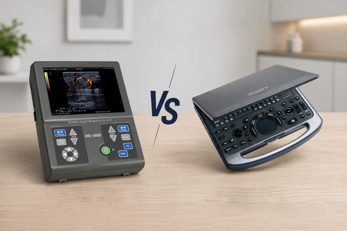
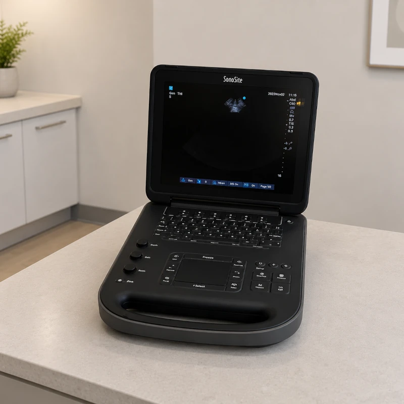
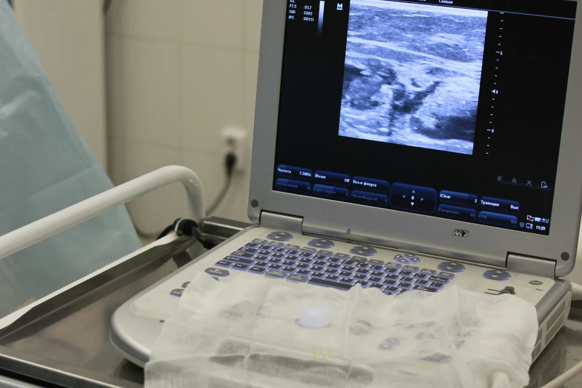
Portable and handheld systems have also come a long way. They may not match every feature of a premium console, but many are now accurate enough for focused bedside or field assessments.
Accuracy depends on the application
Ultrasound tends to be especially reliable when imaging soft tissues and fluid-filled structures.
It works very well for things like the bladder, gallbladder, uterus, fetus, blood vessels, and heart chambers. It is also excellent for detecting free fluid, such as abdominal or pleural effusion.
Where it struggles is with air and bone. Bone reflects most of the ultrasound beam, which creates a bright surface and a shadow behind it. Air scatters the sound waves, which is why gas-filled intestines and lungs can make deeper structures difficult to evaluate. Obesity, patient motion, and deeply located organs can also make imaging harder.
So while ultrasound is highly useful, it is not a universal solution. In some situations, CT, MRI, or radiography may still be the better choice.
Why Ultrasound Accuracy Depends So Much on the Operator
One of the biggest differences between ultrasound and other imaging methods is how operator-dependent it is.
With ultrasound, the person performing the scan has to choose the right probe, adjust the depth and gain, set the focus correctly, angle the transducer properly, and move through the anatomy in a way that reveals the structures of interest. That takes both technical skill and experience.
A skilled operator knows how to:
Position and angle the probe correctly
Even small changes in angle or pressure can dramatically change what is visible.
Adjust settings appropriately
Gain, depth, focus, frequency, and Doppler parameters all need to be tailored to the patient and the question being asked.
Recognize artifacts
Not everything on screen represents pathology. Reverberation, shadowing, and speckle can all be misleading if the operator does not understand them.
Understand anatomy well
Knowing what should be there is essential for recognizing what is abnormal.
Interpret findings in context
Ultrasound does not exist in isolation. It has to be interpreted alongside the patient’s history, physical exam, and other test results.
This is one reason two people using the same machine may not get equally accurate results.
Machine Quality and Probe Selection Matter Too
The machine itself does matter, and so does the transducer.
Frequency and resolution
Higher-frequency probes give better detail, which makes them ideal for superficial structures like tendons, vessels, thyroid tissue, or small animals. The trade-off is that they do not penetrate very deeply.
Lower-frequency probes can image deeper structures, which is helpful for abdominal organs or large patients, but they give less fine detail.
Probe type
Different probes are designed for different tasks.
Linear probes are best for superficial structures and vascular access.
Curvilinear probes are commonly used for abdominal and obstetric imaging because they provide a wider field of view.
Phased-array probes work especially well for cardiac imaging because their small footprint fits between ribs.
Using the wrong probe can make even a good machine seem less accurate than it really is.
Probe condition
A damaged or aging probe can also reduce accuracy. Dead elements, cable faults, or lens damage may cause dropouts or artifacts that affect both image quality and measurement reliability. That is why maintenance and calibration still matter, even with newer systems.
Patient and Animal Factors That Affect Accuracy
Ultrasound does not just depend on the machine and the operator. The patient matters too.
Body size and depth
Larger patients or animals can be harder to scan because the sound waves have to travel farther. That reduces resolution and can make deeper structures less clear.
Gas and bone
Gas in the bowel and bone in the ribs or limbs can block the beam and hide structures behind them. This is one of the biggest practical limitations of ultrasound.
Movement
Motion from breathing, pain, restlessness, or poor restraint can blur the image and make it harder to capture useful views.
Stress or discomfort
A patient that is anxious or painful may not tolerate the positioning needed for a good scan. In veterinary medicine, some animals can be scanned awake, while others may need mild sedation.
Coat, feathers, or scales
In animals, fur, feathers, or scales can reduce contact between the probe and the skin. Shaving and coupling gel are often needed for a useful exam.
Pregnancy stage
In pregnancy imaging, timing matters. Early exams may need different probes and techniques than later ones, and fetal position can also affect what is visible.
Where Ultrasound Is Especially Reliable
Ultrasound is especially strong in several areas.
Pregnancy and reproductive imaging
This is one of the most familiar uses of ultrasound, and for good reason. It is very effective for confirming pregnancy, assessing fetal viability, estimating gestational age, and monitoring growth and movement.
Abdominal imaging
Ultrasound works well for evaluating the liver, kidneys, spleen, bladder, gallbladder, pancreas, and many abdominal masses or fluid collections.
Cardiac imaging
Echocardiography is one of the most valuable applications of ultrasound. It allows clinicians to assess valves, chambers, wall motion, and blood flow in real time.
Vascular imaging
Doppler ultrasound is widely trusted for assessing blood flow in arteries and veins, including thrombosis, stenosis, and abnormal flow patterns.
Guided procedures
Ultrasound is highly reliable for guiding biopsies, fluid aspiration, vascular access, and other procedures where real-time visualization improves safety and precision.
Where Ultrasound Has Limitations
Even today, ultrasound still has some clear limits.
Bone and air remain major obstacles
Structures hidden behind ribs, skull, or gas-filled bowel can be difficult or impossible to evaluate fully.
Deep anatomy is harder to image
Although low-frequency probes help with depth, the image becomes less detailed the deeper you go.
Limited field of view
Ultrasound shows a slice of anatomy rather than the larger overview you might get with CT or MRI.
Artifacts can still mislead
Reverberation, shadowing, and other artifacts remain part of ultrasound and can either mimic disease or hide it.
It depends heavily on the user
This is still one of ultrasound’s defining limitations. Technology helps, but it does not eliminate the need for training and experience.
Are Newer Ultrasound Machines More Accurate Than Older Ones?
In general, yes, newer ultrasound systems are usually more accurate and easier to use than older ones.
They often offer better image resolution, faster processing, more advanced Doppler features, stronger ergonomics, automated measurements, and newer imaging modes like elastography or 3D and 4D imaging. Portable systems have improved dramatically too.
That said, an older machine that is well maintained and used by an experienced operator can still perform very well. In many cases, probe condition, proper settings, and user skill matter just as much as the age of the platform.
How AI Is Improving Ultrasound Accuracy
Artificial intelligence is starting to play a bigger role in ultrasound, especially in helping reduce variability.
AI can help by:
Automating measurements
This reduces manual variability and saves time.
Improving image acquisition
Some systems can guide the user toward better probe positioning or settings.
Recognizing anatomy
AI tools can label structures or help identify standard planes.
Supporting quality control
Some systems can flag poor images or possible transducer performance issues.
Providing decision support
In some cases, AI may suggest patterns or findings that help the clinician.
AI can improve consistency, especially for less experienced users, but it does not replace a trained sonographer, veterinarian, or physician. It is a support tool, not a substitute for judgment.
How to Improve Ultrasound Accuracy in Practice
A few practical steps can make a big difference in everyday use.
Choose the right probe
Match the probe type and frequency to the anatomy being scanned.
Optimize the settings
Depth, gain, focus, and Doppler settings should be adjusted for each exam.
Maintain the equipment
Probe checks, calibration, and preventive maintenance all help preserve performance.
Invest in training
Ultrasound skill improves with education, repetition, and review.
Position the patient well
Good positioning and gentle restraint reduce motion and improve image quality.
Use ultrasound in context
Findings should always be considered alongside history, physical exam, lab work, and other imaging when needed.
Review and document studies
Saving images and reviewing cases helps improve consistency and quality over time.
Conclusion
Ultrasound machines today are more advanced, more portable, and more capable than ever before. They provide high-quality real-time imaging without radiation and are especially reliable for soft tissue, vascular, cardiac, and reproductive imaging.
But ultrasound accuracy is still shaped by more than just the machine. The best results come from the right probe, the right settings, careful maintenance, good patient handling, and a skilled operator who understands both the technology and the anatomy.
So, how accurate are ultrasound machines today? In the right hands and for the right applications, they can be extremely accurate. The key is knowing both what ultrasound does well and where its limits still are.
FAQ
How accurate are ultrasound machines today?
They can be very accurate for many soft tissue, vascular, cardiac, and pregnancy-related exams. The exact accuracy depends on the type of exam, the machine, the probe, the patient, and the operator.
Are newer ultrasound machines more accurate than older ones?
Usually, yes. Newer systems often have better image resolution, improved Doppler, faster processing, and more advanced software. Still, a well-maintained older system can also perform very well in experienced hands.
What affects ultrasound accuracy the most?
Operator skill is one of the biggest factors. Probe selection, machine quality, settings, patient anatomy, motion, and the specific condition being evaluated also play major roles.
Is ultrasound more accurate than X-ray?
Not generally better overall, just better for different things. Ultrasound is stronger for soft tissues, blood flow, pregnancy, and real-time imaging. X-rays are better for bones, air-filled lungs, and certain foreign objects.
Can ultrasound results be wrong?
Yes. Poor image windows, gas, bone, motion, incorrect settings, damaged probes, or limited operator experience can all reduce accuracy or lead to misinterpretation.
Does AI make ultrasound more accurate?
AI can improve consistency by helping with measurements, image optimization, and anatomy recognition. It can support accuracy, but it does not replace trained professionals.
Are portable ultrasound machines accurate?
Many are accurate enough for focused exams and point-of-care use, especially when paired with the right probe and used by someone trained. They may still have fewer features than premium cart-based systems.
How accurate is ultrasound for pregnancy?
Ultrasound is widely considered very accurate for confirming pregnancy, checking fetal viability, estimating gestational age, and monitoring development.
How can clinics improve ultrasound accuracy?
By using the right probes, maintaining equipment, training operators well, optimizing settings, positioning patients properly, and interpreting findings alongside other clinical information.
Does ultrasound use radiation?
No. Diagnostic ultrasound uses high-frequency sound waves, not ionizing radiation.